 Corporate Website
 Africa
 Argentina
 Asia
Asia
 Australia
 Belgium
 Brazil
 Bulgaria
 Canada (EN)
 Chile
Chile
 China
China
 Colombia
 Denmark
 Egypt
Egypt
 France
 Germany
 Greece
 Hungary
 Indonesia
 Italia
 India
India
 Japan
Japan
 Korea
Korea
 Malaysia
 Mexico
 Middle East
 Netherlands
 Peru
Peru
 Philippines
 Poland
 Portugal
 Romania
 Russia
 South Africa
 Spain
Spain
 Sweden
 Thailand
 Tunisia
 Turkey
 Ukraine
 United Kingdom
 USA
USA
 Vietnam
Mycoplasma hyopneumoniae is the primary pathogen causing Enzootic Pneumonia (EP), one of the most prevalent respiratory diseases in pigs resulting in high morbidity but low mortality. It is also one of the main pathogens involved in the porcine respiratory disease complex (PRDC), however other bacteria and viruses are also often contributing factors.
Acute inflammation, due to Mycoplasma hyopneumoniae infection, leads to a catarrhal or even purulent exudate in the airways. The accumulation of neutrophils and mono-nuclear cells around the bronchi and bronchioles results in airway collapse and consolidation of the lungs.
Figure 1: Pig lungs - Lesions of broncho-pneumonia and cranial pleurisy*
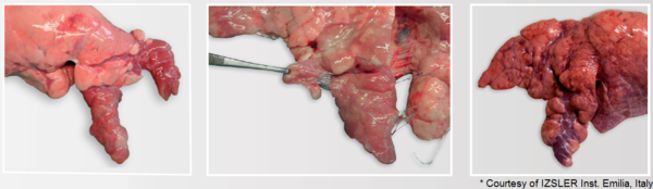
Lung tissue infected with Mycoplasma hyopneumoniae develops consolidation and catarrhal broncho-pneumonia with purple to grey regions of meaty appearance. The consolidation can be observed from 3 - 12 weeks post infection. The lesions are mainly localized in the apical and cardiac lobes, in the anterior part of the diaphragmatic lobes, as well as in the intermediate lobe. Lesions resolve after 12 to 14 weeks with formation of interlobular fissures, also referred to as scarring.
Mycoplasma hyopneumoniae and related viral and bacterial pathogens are also responsible for the development of pleuritis, which is typically localized in the cranio-ventral part of the lungs, most often between the apical and cardiac lobes.
Consolidation of the apical and cardiac lung lobes indicating the acute to sub-acute bronchopneumonia is suggestive of Mycoplasma hyopneumoniae induced EP. Pleuritis at the junction of the cardiac and diaphragmatic lobes can still be assigned to the cranio-ventral area. Pleurisy in this area are most likely due to Mycoplasma hyopneumoniae and associated bacterial or viral infections.
<< Back to Lung Scoring Methodology
Related topics: mycoplasma hyopneumoniae ceva lung program swine lung scoring ep enzootic pneumonia


