 Corporate Website
 Africa
 Argentina
 Asia
Asia
 Australia
 Belgium
 Brazil
 Bulgaria
 Canada (EN)
 Chile
Chile
 China
China
 Colombia
 Denmark
 Egypt
Egypt
 France
 Germany
 Greece
 Hungary
 Indonesia
 Italia
 India
India
 Japan
Japan
 Korea
Korea
 Malaysia
 Mexico
 Middle East
 Netherlands
 Peru
Peru
 Philippines
 Poland
 Portugal
 Romania
 Russia
 South Africa
 Spain
Spain
 Sweden
 Thailand
 Tunisia
 Turkey
 Ukraine
 United Kingdom
 USA
USA
 Vietnam
--> Introduction
--> Results
--> Discussion and Conclusions
.
TISSUE CONCENTRATIONS OF AMOXICILLIN IN SUCKLING PIGLETS AFTER SINGLE INJECTION OF VETRIMOXIN® LA
By Krejci R.1, Guyonnet J.1, Robert N.1, Malasek J.2, Honzlova A.3
1Ceva, Libourne, France; 2Veterinary consultant, Zdar n Sazavou, Czech Rep.; 3State Veterinary Institute, Jihlava, Czech Rep.
.
Introduction
Amoxicillin is one of the most frequently used antibiotics in pigs for the treatment and prophylaxis of various pathologies. The long acting injectable forms allow controlled individual treatment of sick animals or animals at the imminent risk of infection.
Suckling piglets in industrial farms are exposed to highly contagious early colonizers such as Streptococcus suis (S. suis) or Haemophilus parasuis (H. parasuis), invasive bacteria able to spread quickly into various tissues of the infected individual. The blood concentrations of amoxicillin, which is the antibiotic of choice in the control of such infections, were well documented after a single injection of the long acting preparations. There is, however very limited knowledge about the concentrations in tissues.
.
Materials and Methods
Healthy suckling piglets of the weight about 4 kg were injected once with the doses of either 0.1ml per kg lbw or 0.5ml per kg lbw with Vetrimoxin® LA, Ceva (150mg amoxicillin trihydrate in 1 ml). Five piglets were included in each dose group.
Animals were euthanized 2 hours later which corresponds to the Tmax previously assessed by the PK study with Vetrimoxin® LA in 30kg pigs. Blood, synovial fluid and tissues of brain, tonsils and lungs were collected at this time from each of tested piglets. Samples were examined using HLPC/MS/MS method for the quantifiation of amoxicillin.
.
Results
The results revealed the average concentrations in the brain, lungs, tonsils, blood and synovial flid: 0.08 μg/g, 0.06 μg/g, 0.08 μg/g, 4.12 μg/ml and 0.03 μg/ml respectively after the dose of 0.1ml.kg lbw (Figure 1) and 0.12 μg/g, 0.20 μg/g, 0.22 μg/g, 11.25 μg/g and 0.15 μg/g respectively after the dose of 0.5ml/kg lbw (Figure 2).
Figure 1 Plasma and tissue concentrations determined 2 hours after the i.m. dose of 15mg/kg
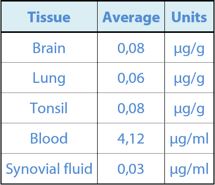
The ratio tissue/plasma ranged between 0.0774 and 0.019, with the highest ratios found in the tonsils and the brain.
Figure 2 Plasma and tissue concentrations determined 2 hours after the i.m. dose of 75mg/kg
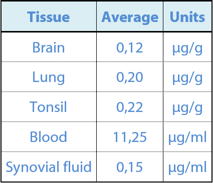
The highest ratios tissue/plasma were found in the tonsils and lungs, 0.19 and 0.18 respectively. The plasma concentrations were approximately 3 times higher than the ones achieved after the dose of 15mg/kg, which was 5 times higher.
.
Discussion and Conclusions
Concentrations in lungs, brain, synovial flid and lymphoid tissue observed in this study in suckling piglets were approximately within the same rank of values as reported by Agerso et al. (1998). It means that, for instance the period for which the concentration in the synovial fluid is above the MIC for S. suis (0.03 mg/ml, Ceva internal data) lasts about 48 hours after the dose of 15mg/kg.
After the dose of 75 mg/kg (5 x 15 mg/kg), the concentrations measured 2 hours post injection were three times higher than after the dose of 15 mg/kg and did not reach the 5 times equivalent. This is explained by the delay to achieve the Cmax, which also indicates longer duration of the concentrations above the MIC. Further studies are needed to set the full profie of the concentrations in the tissues.
.
References
Agerso H. et al., 1998, Res. Vet. Sci., 64, 251-257
(Source: Axis Issue 02 / Sept 2013 - Ceva Asia Pacific - Proceedings APVS 2013)
.


