 Corporate Website
 Africa
 Argentina
 Asia
Asia
 Australia
 Belgium
 Brazil
 Bulgaria
 Canada (EN)
 Chile
Chile
 China
China
 Colombia
 Denmark
 Egypt
Egypt
 France
 Germany
 Greece
 Hungary
 Indonesia
 Italia
 India
India
 Japan
Japan
 Korea
Korea
 Malaysia
 Mexico
 Middle East
 Netherlands
 Peru
Peru
 Philippines
 Poland
 Portugal
 Romania
 Russia
 South Africa
 Spain
Spain
 Sweden
 Thailand
 Tunisia
 Turkey
 Ukraine
 United Kingdom
 USA
USA
 Vietnam
Avian salmonelloses could be classified in two groups. The first one include infections (pullorum disease and fowl typhoid) caused by the two non-motile serotype S. pullorum and S.gallinarum. The second group comprises infections caused by multiple motile Salmonella serotypes - most frequently S. Enteritidis and S. Typhimurium isolates that are considered together as paratyphoid.
PULLORUM DISEASE
Pullorum disease is an acute systemic disease in chickens and turkey poults. The infection is transmitted with eggs and is commonly characterized by a white diarrhoea (Image 1) and high death rate, whereas adult birds are asymptomatic carriers. The morbidity and the mortality rates increase about the 7th - 10th day after hatching. The affected chickens appear somnolent, depressed and their growth is retarded. The feathers around the vent in many chickens is stained with diarrhoeic faeces or pasted with dry faeces (Image 2).
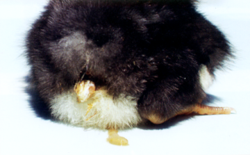
Image 1 White diarrhoea in the chickens poult
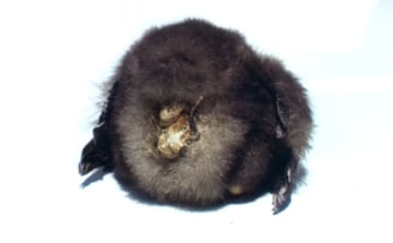
Image 2 The dry diarrhoeic faeces in the vent
The oedema of tibiotarsal joints is a frequent associated sign (Image 3). Pullorum disease is widely distributed among all age groups of chickens and turkeys. The highest losses are in birds under the age of 4 weeks.
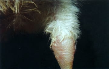
Image 3 The oedema of tibiotarsal joints
The aetiological agent is S. pullorum, a non-motile Gram-negative micro-organism. S. pullorum is very resistant under moderate climatic conditions and could survive for months. It could be killed by fumigation with formaldehyde of breeder eggs in the hatchery.
Typical for this form are the greyish-whitish nodes in one or some of the following places: heart (Image 4), lungs, liver, gizzard walls (Image 5) and intestines, the peritoneum.
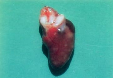
Image 4 The greyish-whitish nodes in the heart
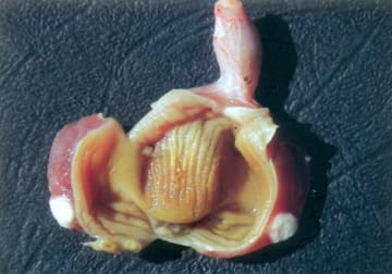
Image 5 The greyish-whitish nodes in the gizzard walls
Sometimes, greyish-whitish milliary necroses are found out in the liver (Image 6). S. pullorum is transmitted by infected eggs of layer hens that are carriers. Many hatched infected chickens spread the micro-organism by a horizontal route to other birds via the gastro-intestinal and the urinary tracts. Adult carrier birds also spread the agent through their excreta.
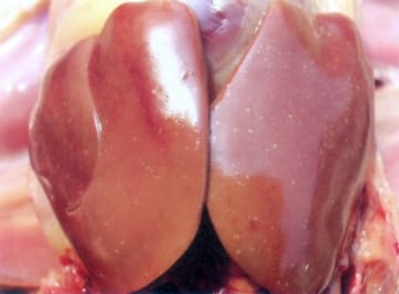
Image 6 Greyish-whitish milliary necroses in the liver
Enlarged and septicaemic spleen (Image 7).
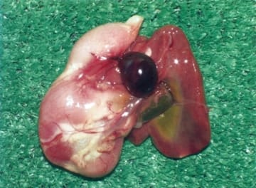
Image 7 Enlarged and septicaemic spleen
For confirmation of the diagnosis, S. pullorum should be isolated and typed.
Pullorum disease must be differentiated from other salmonelloses, E. coli infections, Aspergillus that produces similar pulmonary lesions, Staphylococcus aureus caussing arthrites, etc. Sometimes, the pulmonary nodes resemble the tumours in Marek’s disease.
FOWL TYPHOID
Fowl Typhoid is an acute or chronic septicaemic disease that affects primarily adult hens and turkeys.
The aetiological agent is Salmonella gallinarum. This organism usually shares common antigens with S. pullorum and the two micro-organisms often give a cross-agglutination reaction.
The transmission of the infection by contaminated eggs is especially important. Moreover, the transmission of S. gallinarum occurs mainly among growing or productive flocks and the death rate among adult birds is higher.
Acute fowl typhoid
The outbreaks usually begin with a sharp decline in forage consumption and egg production. The fertilization and hatchability rates are considerably reduced. Diarrhoea appears. The death rate in acute fowl typhoid is high and varies between 10% and 90%. About 1/3 of chickens hatched from eggs from typhoid-infected flocks die. A characteristic lesion for acute fowl typhoid in adult birds is the enlarged and bronze greenish tint of liver (Image 8).
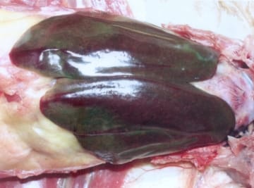
Image 8 Enlarged and bronze greenish tint of liver
In some instances, the enlarged liver is mottled with multiple miliary necroses (Image 9). The outbreaks are observed primarily in hens and turkeys, but the diseases is sometimes encountered in other domestic or wild fowl.
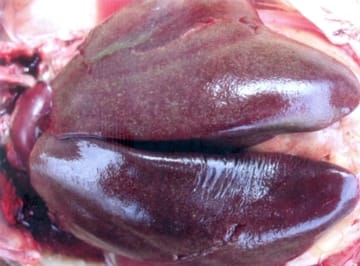
Image 9 The multiple miliary necroses in the enlarged liver
In other cases, the size of liver necroses varies from miliary to spots with a diameter of 1 - 2 cm (Image 10). Unlike pullorum disease, fowl typhoid is lasting for months.
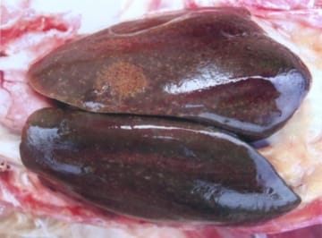
Image 10 The spot necroses in the liver with a diameter of 1 - 2 cm
The spleen is 2 - 3 times bigger, sometimes with greyish-whitish nodules prominating on the surface, representing hyperplastic follicles (Image 11).
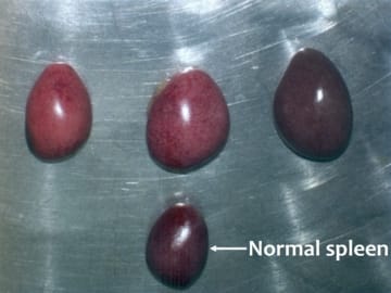
Image 11 The spleen is 2 - 3 times bigger with greyish-whitish nodules prominating on the surface
Often, enteritis, especially of the anterior part of small intestine, sometimes with ulcerations, is present (Image 12).
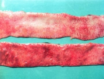
Image 12 Enteritis with ulcerations
More rarely, myocardial necroses due to Salmonella toxins are detected (Image 13).
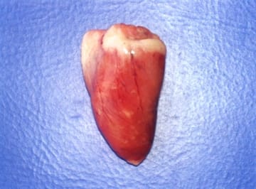
Image 13 Myocardial necroses
The lungs acquire a characteristic brown colour (Image 14). Here, necroses and, following their organization, “sarcoma-like nodules” could be observed.
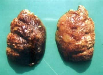
Image 14 The brown colour of the lungs with "sarcoma-like nodules”
Chronic fowl typhoid
The lesions are primarily in the gonads. The ovaries are affected by inflammatory and degenerative changes (Image 15).
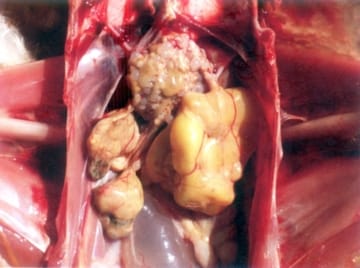
Image 15 The inflammatory and degenerative ovaries
Frequently, affected follicles are deformed and appear like thick pendulating masses (Image 16).
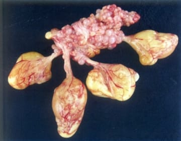
Image 16 The deformed follicles with thick pendulating masses
Taking into consideration that chemotherapy does not eliminate the carriership, the treatment of poultry infected with fowl typhoid or pullorum disease is not justified and is never recommended.
If breeder flocks are proved to be carriers of the infection, their eggs should not be used for breeding.
Fowl typhoid should be differentiated from other salmonelloses, E. coli infections, Pasteurella spp. infections, etc.
PARATYPHOID INFECTIONS
Fowl paratyphoid is an acute or chronic disease in domestic fowl and many other avian or mammalian species, caused by some motile Salmonella serotypes that are not host-specific. The highest morbidity and death rates are usually observed during the first 2 weeks after hatching. Haemorrhagic fibrinous typhlitis is a possible finding (Image 17). Most fowl paratyphoid organisms contain an endotoxin, responsible for their pathogenic effects. Diarrhoea, dehydration and pasted down appearance around the vent are observed.
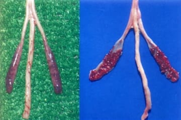
Image 17 Haemorrhagic fibrinous typhlitis
The aetiological agents are about 10 - 15 Salmonella serotypes and the most common isolates are S. Enteritidis and S. Typhimurium. Pathoanatomically, marked catarrhal haemorrhagic enteritis is observed. Often the caeca are filled with gelatinous, fibrinous, cheese-like exudate. This is a finding, characteristic for salmonellosis, but it is not specific for any of serotypes. The inflammatory fibrinous exudate in caeca often forms casts with the shape of mucosal folds (Image 18).
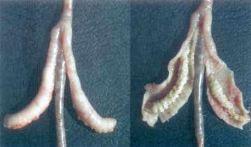
Image 18 The inflammatory fibrinous exudate in caeca forms casts with the shape of mucosal folds
Sometimes, necrotic foci in the liver are discovered (Image 19).
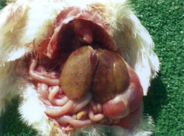
Image 19 Necrotic foci in the liver
The infection of small chickens occurs by penetration of micro-organisms into the egg after faecal contamination. The transmission of agents could be done also by a contaminated source of animal protein (meat and bone meal, etc.). The rodents are a significant reservoir of paratyphoid micro-organisms.
The treatment inhibits but does not eradicate the infection. The appropriate treatment minimizes the death rate until the birds develop immunity.
(Source: "Diseases of poultry - A colour atlas" - Ivan Dinev & CEVA Santé Animal, 2010)
.
Related topics: se st disease information technical poultry salmonella salmonellose


