 Corporate Website
 Africa
 Argentina
 Asia
Asia
 Australia
 Belgium
 Brazil
 Bulgaria
 Canada (EN)
 Chile
Chile
 China
China
 Colombia
 Denmark
 Egypt
Egypt
 France
 Germany
 Greece
 Hungary
 Indonesia
 Italia
 India
India
 Japan
Japan
 Korea
Korea
 Malaysia
 Mexico
 Middle East
 Netherlands
 Peru
Peru
 Philippines
 Poland
 Portugal
 Romania
 Russia
 South Africa
 Spain
Spain
 Sweden
 Thailand
 Tunisia
 Turkey
 Ukraine
 United Kingdom
 USA
USA
 Vietnam
The Newcastle disease (ND) is a highly contagious disease in many species of domestic, exotic and wild birds that, depending on its tropism, is characterized by marked variations in morbidity, death rate, symptoms and lesions. It is caused by a paramyxovirus. Birds at any age are susceptible. The clinico-morphological signs possess a distinct viscerotropic or neurotropic character.
In the viscerotropic form, haemorrhagic diphtheritic lesions of the entire alimentary tract, from the break to the vent, are present. The haemorrhages of gizzard epithelium are remarkable (Image 1). The mucous coat is oedematous, covered with thick mucus (Image 2) and mottled with haemorrhages varying from single to multiple (Image 3), sometimes gathered at the boundaries with the gizzard or the oesophagus.
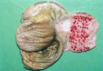
Image 1 The haemorrhages of gizzard epithelium
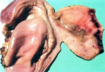
Image 2 The mucous coat of gizzard is oedematous, covered with thick mucus
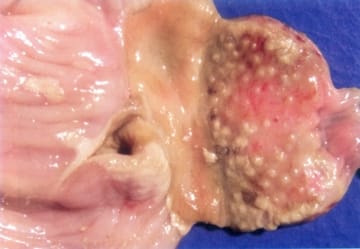
Image 3 The mucous coat of gizzard is mottled with haemorrhages
Typical for this form are the haemorrhagic necrotic and focal diphtheroid lesions affecting the mucosa of the buccal cavity (Image 4), the stomach and the intestines (Image 5). The disease is generally prevalent in hens, more rarely in turkeys, exotic or wild birds.
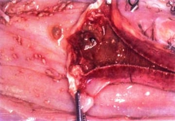
Image 4 The haemorrhagic necrotic and focal diphtheroid lesions in the mucosa of the buccal cavity
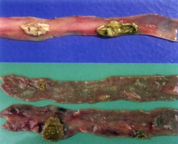
Image 5 The haemorrhagic necrotic and focal diphtheroid lesions in the mucosa of the stomach and the intestines
Depending on their pathogenicity in chicken embryos, the numerous known strains are classified as lentogenic, mesogenic and velogenic. The vaccines made of lentogenic strains provoke a shorter immunity that requires a revaccination. The vaccines from mesogenic strains result in a lasting immunity, but could provoke a lethal issue especially in birds without a primary immunity created on the basis of lentogenic vaccinal strains.
A frequent finding is the enlargement and haemorrhages of caecal tonsils (Image 6) and haemorrhagic cloacitis (Image 7). Usually, these lesions begin from the lymphoid tissue of the mucous coat.
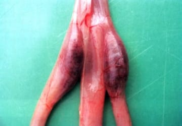
Image 6 The enlargement and haemorrhages of caecal tonsils
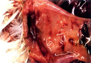
Image 7 Haemorrhagic cloacitis
The virus, contained in incubated eggs, results in embryo’s death and then perishes.
Virus-containing excreta of infected birds, that contaminate the forage, water and the environment, are the source of infection. The infection is transmitted mainly by an oral route, the airborne or contact transmission being more infrequent. There is no permanent carriership of the virus. An important factor in the transmission of velogenic virues could be the exotic birds and fighting cocks. The death rate could arrive at 70 - 100%.
The neurotropic form of the disease is clinically manifested by ataxia, opisthotonus (Image 8), torticolis, paresis and paralysis of legs. This form is frequently accompanied by respiratory symptoms. Histopathologically, the picture of nonpurulent lymphocytic encephalomyelitis is observed.
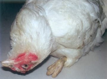
Image 8 Opisthotonus in the neurotropic form
(Source: "Diseases of poultry - A colour atlas" - Ivan Dinev & CEVA Santé Animal, 2010)
.


