 Corporate Website
 Africa
 Argentina
 Asia
Asia
 Australia
 Belgium
 Brazil
 Bulgaria
 Canada (EN)
 Chile
Chile
 China
China
 Colombia
 Denmark
 Egypt
Egypt
 France
 Germany
 Greece
 Hungary
 Indonesia
 Italia
 India
India
 Japan
Japan
 Korea
Korea
 Malaysia
 Mexico
 Middle East
 Netherlands
 Peru
Peru
 Philippines
 Poland
 Portugal
 Romania
 Russia
 South Africa
 Spain
Spain
 Sweden
 Thailand
 Tunisia
 Turkey
 Ukraine
 United Kingdom
 USA
USA
 Vietnam
IBD is an acute, highly contagious viral infection in chickens manifested by inlammation and subsequent atrophy of the bursa of Fabricius, various degrees of nephrosonephritis and immunosuppression. Clinically the disease is seen only in chickens older than 3 weeks. The feathers around the vent are usually stained with faeces containing plenty of urates (Image 1).
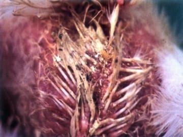
Image 1 The feathers around the vent are stained with faeces containing plenty of urates
The period of most apparent clinical symptoms and high death rate is at the age of 3 - 6 weeks. IBD could however be observed as long as chickens have a functioning bursa (up to the age of 16 weeks). In chickens younger than 3 weeks, IBD could be subclinical, but injured bursa leads to immunosuppression. Also, diarrhoea, anorexia, depression, ruffled feathers, especially in the region of the head and the neck are present (Image 2).
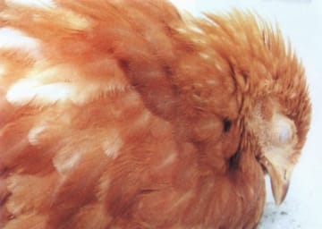
Image 2 Chicken is depressed and has the ruffled feathers
A natural IBD infection is mostly observed in chickens. In turkeys and ducks it could occur subclinically, without immunosuppression. Most isolates of the IBD virus in turkeys are serologically different from those in chickens. In premises, once contaminated with the IBD virus, the disease tends to recur, usually as subclinical infection. The dead bodies are dehydrated, often with haemorrhages in the pectoral, thigh and abdominal muscles (Image 3).
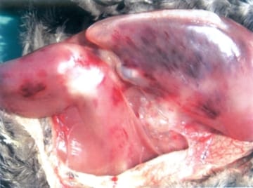
Image 3 Hemorrhages in the pectoral, thigh and abdominal muscle
The IBD virus belongs to the Birnaviridae family of RNA viruses. Two serotypes are known to exist, but only serotype 1 is pathogenic.
The virus is highly resistant to most disinfectants and environmental conditions. In contaminated premises, it could persist for months and in water, forage and faeces - for weeks.
The incubation period is short and the first symptoms appear 2 - 3 days after infection. The lesions in the bursa of Fabricius are progressive. In the beginning, the bursa is enlarged, oedematous and covered with a gelatinous transudate (Image 4).
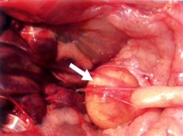
Image 4 The bursa is covered with a gelatinous transudate
The IBD virus has a lymphocidic effect and the most severe injuries are in the lymph follicles of the bursa of Fabricius. Most commonly, IBD begins as a serous bursitis (Image 5).
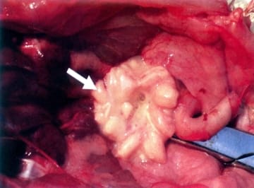
Image 5 A serous bursitis
IBD lesions undergo various stages of serous haemorrhagic to severe haemorrhagic inflammation (Image 6).
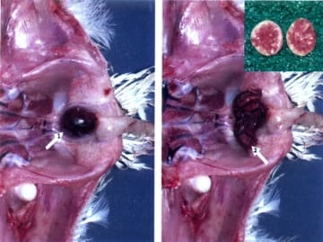
Image 6 Serous haemorrhagic (left) and severe haemorrhagic inflammation (right)
The morbidity rate is very high and could reach 100%, whereas the mortality rate: 20 - 30%. The course of the disease is 5 - 7 days and the peak mortality occurs in the middle of this period.
In some cases, the bursa is filled with coagulated fibrinous exudate that usually forms casts with the shape of mucosal folds (Image 7).
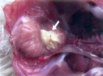
Image 7 Coagulated fibrinous exudate forms casts with the shape of mucosal folds of the bursa
In birds surviving the acute stage of the disease, the bursa is progressively atrophying. Microscopically, an atrophy of follicles into the bursa is observed secondary to inflammatory and dystrophic necrobiotic alterations.
The kidneys are affected by a severe urate diathesis (Image 8).
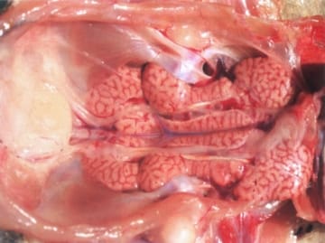
Image 8 The kidneys are affected by a severe urate diathesis
In an acute outbreak and manifestation of the typical clinical signs, the diagnostics is not difficult. The diagnosis could be confirmed by detection of typical gross lesions throughout a pathoanatomical study.
IBD should be differentiated from IBH (inclusion body hepatitis).
The application of live vaccines in chickens is a key point in the prevention of IBD and should be related to the levels of maternal antibodies.
(Source: "Diseases of poultry - A colour atlas" - Ivan Dinev & CEVA Santé Animal, 2010)
.
Related topics: fabricius disease information technical poutry ibd infectious bursa gumboro


